“I believe we can ensure the facility continues to empower innovative, world-class science – while remaining accessible, flexible, and researcher-focused”
Histology is the microscopic study of tissues and organs through sectioning, staining, and examining those sections under a microscope.
The Research Histology Facility is a key component of the Inflammation, Repair and Development (IRD) Section at the National Heart and Lung Institute (NHLI), providing essential services such as paraffin processing and embedding and cryosectioning whilst also offering advice on protocol development and troubleshooting for immunohistochemistry staining.
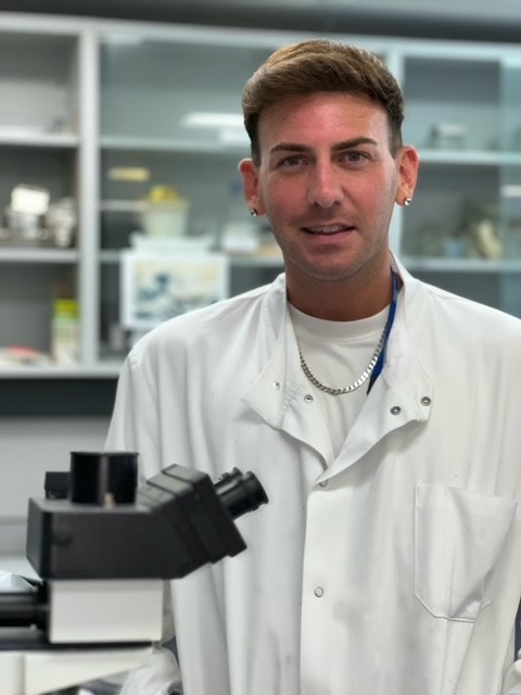
We had the opportunity to speak with Connor Preston, Senior Technician overseeing the IRD Histology Facility, to learn about his day-to-day responsibilities, his passion for discovery, and how his role aligns with his goals of fostering stronger interdisciplinary collaborations within the facility.
Can you tell us about your role at the Research Histology Facility? What does a typical day look like for you?
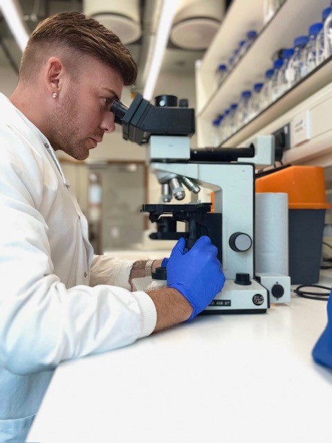
As the Senior Technician overseeing the IRD Histology Facility at the National Heart & Lung Institute, I provide a full histological service to support researchers across the NHLI and wider Imperial community. On any given day, I could be processing samples, embedding tissue, cryosectioning, performing stains (H&E, special stains, IHC/IF), or troubleshooting protocols for researchers.
Alongside the hands-on lab work, I manage the daily operations of the facility, coordinating bookings, ordering reagents, maintaining equipment, ensuring compliance with COSHH and HTA regulations, and training PhD students and postdocs. It’s a role that blends technical precision with people-focused collaboration, and no two days are the same – which is what I love about it.
How did you first get involved in Histology, and what attracted you to this field?
My journey into histology began in clinical histopathology at Great Ormond Street Hospital. I was fascinated by the way tissue preparation and staining could reveal so much about disease at a cellular level. That curiosity led me to explore research histology, which offered more scope for discovery and experimental development.
Over the years, I’ve worked in varied environments – from diagnostic services to world-leading academic institutions like UCL and Imperial – but the common thread has always been the excitement of uncovering the microscopic stories within tissues, and helping researchers find answers to complex biological questions. It is endlessly fascinating.
What are some of the key techniques and technologies used in the Histology Facility, and how do they support research?
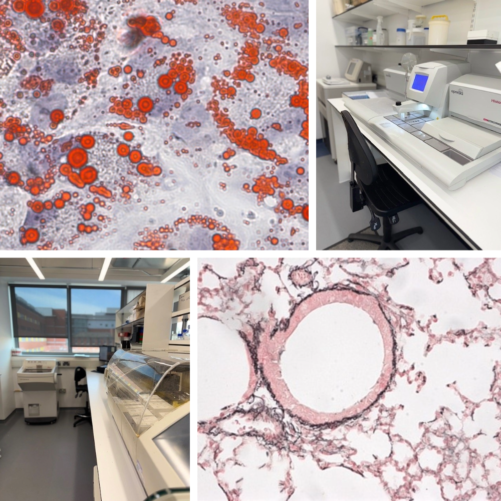
We provide a broad range of histological techniques, including:
- Paraffin processing and embedding
- Cryosectioning and paraffin microtomy
- H&E, special stains (e.g. Masson’s Trichrome, Alcian Blue), and enzyme histochemistry
- Immunohistochemistry and immunofluorescence
- Imaging via brightfield and fluorescence microscopy
These techniques are critical for visualising tissue architecture, identifying cell types, and localising proteins or pathological changes. For example, in respiratory research, we can assess inflammation, fibrosis, or airway remodelling in lung tissue, while in cardiovascular studies, we might look at cardiac fibrosis or vessel changes.
Our facility allows researchers to integrate morphological and molecular data, strengthening their findings and adding visual depth to their studies.
“It’s a role that blends technical precision with people-focused collaboration, and no two days are the same – which is what I love about it”
How does your work directly impact the research conducted at Imperial, particularly in areas like cardiovascular, respiratory, and other medical research?
Our histology work underpins a wide range of NHLI research, including studies on asthma, COPD, fibrosis, cardiovascular disease, and regenerative medicine. By offering a reliable and flexible histological service, we give researchers access to high-quality tissue data that is essential for interpreting results and publishing impactful science.
For example, we support the visual validation of experimental models – whether that’s showing immune infiltration in lung sections or identifying scar formation post-myocardial infarction. My background in both clinical and research histology helps bridge the gap between basic research and translational outcomes, ensuring that tissue-based data is accurate, reproducible, and publication-ready.
Can you share a success story or breakthrough for the Histology Facility?
A recent highlight has been my involvement in projects using Xenium In Situ spatial transcriptomics, a cutting-edge platform that allows researchers to detect and map RNA transcripts at subcellular resolution in complex tissues like the lung or heart.
This project has not only expanded the capabilities of our facility but has also positioned us to support more transcriptomics-based research in the future.
“Histology underpins so much of biomedical research, and I see the facility becoming a central hub for integrated tissue-based approaches”
What are your goals for the facility, and how do you envision its growth and impact in the future?
Looking ahead, my goals include:
- Expanding our digital pathology and image analysis capabilities
- Introducing spatial transcriptomics workflows
- Offering more tailored training sessions for students and early-career researchers
A key priority is growing the Histology Facility team. With increased staffing, we’d be able to provide a more consistent, high-throughput service, maintaining the high quality and reliability our users depend on. A larger team would also allow us to take on more complex or longitudinal projects without affecting routine services.
I’m especially keen to build stronger interdisciplinary collaborations – both within NHLI and across departments at Imperial. Histology underpins so much of biomedical research, and I see the facility becoming a central hub for integrated tissue-based approaches, from traditional staining to advanced spatial transcriptomic techniques.
By expanding our capacity, modernising our services, and deepening research partnerships, I believe we can ensure the facility continues to empower innovative, world-class science – while remaining accessible, flexible, and researcher-focused.
Find out more about the Histology facility and other facilities available at NHLI.