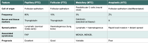
Blog posts

Duodenal biopsy site for diagnosing coeliac disease: D1, D2, or both?

Summary

Current best practice, in line with BSG, ESPGHAN and ACG guidance, is to take biopsies from both the duodenal bulb (D1) and the distal duodenum (D2 / D3), placed in separate pots. At least one (preferably two) bulb biopsies plus four or more distal duodenal biopsies is the standard recommendation. D2 remains the principal diagnostic site, but adding D1 increases diagnostic sensitivity, particularly for ultra-short coeliac disease and paediatric cases.

Historical position: D2 only

Traditional teaching was to biopsy the second part of the duodenum and to avoid the bulb. The rationale was that the bulb is architecturally complex — Brunner glands distort villous architecture, peptic injury and gastric metaplasia are common, and orientation in the histology laboratory is often poor. These features were felt to give unreliable assessment of villous atrophy and to risk false-positive interpretations of Marsh 1–3 change.

Why D1 was added: ultra-short coeliac disease

A series of studies in the 2000s and 2010s showed that a meaningful minority of coeliac patients have disease that is confined to, or most marked in, the bulb. Bonamico and colleagues demonstrated that taking bulb biopsies in addition to distal duodenum increased the diagnostic yield in children, and identified the 9 o’clock and 12 o’clock positions as the most informative bulb sites. Evans et al. and subsequent adult series have confirmed that around 5–10% of adult coeliac patients have lesions limited to or more severe in the bulb (ultra-short coeliac disease), and that omitting D1 would miss these cases.

Practical protocol

  • At least 1–2 biopsies from the duodenal bulb (ideally from the 9 o’clock and 12 o’clock positions, per Bonamico).
  • At least 4 biopsies from the distal duodenum (D2 / D3).
  • Bulb biopsies should be submitted in a separate, clearly labelled pot so the pathologist can apply appropriate caution when interpreting villous architecture around Brunner glands.
  • Single-biopsy-per-pass technique is preferred for orientation; multiple biopsies in one bite increase tangential sectioning and false-positive villous blunting.

Caveats with bulb biopsies

Bulb mucosa is intrinsically harder to assess. Brunner gland lobules push villi apart and shorten them, gastric metaplasia is common, and peptic duodenitis can produce IEL increases unrelated to gluten. Bulb-only abnormalities should therefore be interpreted in the context of serology, HLA status, and the distal duodenal appearances before a confident coeliac diagnosis is made.

Bottom line

Biopsy both. D2 remains the principal diagnostic site, but the addition of separately-potted bulb biopsies materially improves sensitivity, particularly for ultra-short coeliac disease and in children. This is the position of the BSG, ESPGHAN and ACG guidelines.

Key references

  • Ludvigsson JF, Bai JC, Biagi F, et al. Diagnosis and management of adult coeliac disease: guidelines from the British Society of Gastroenterology. Gut 2014;63:1210–1228.
  • Husby S, Koletzko S, Korponay-Szabó I, et al. European Society Paediatric Gastroenterology, Hepatology and Nutrition Guidelines for Diagnosing Coeliac Disease 2020. J Pediatr Gastroenterol Nutr 2020;70:141–156.
  • Rubio-Tapia A, Hill ID, Kelly CP, et al. ACG Clinical Guidelines: Diagnosis and Management of Celiac Disease. Am J Gastroenterol 2013;108:656–676 (and updated 2023 ACG guideline, Am J Gastroenterol 2023;118:59–76).
  • Bonamico M, Mariani P, Thanasi E, et al. Patchy villous atrophy of the duodenum in childhood celiac disease. J Pediatr Gastroenterol Nutr 2004;38:204–207.
  • Bonamico M, Thanasi E, Mariani P, et al. Duodenal bulb biopsies in celiac disease: a multicenter study. J Pediatr Gastroenterol Nutr 2008;47:618–622.
  • Evans KE, Aziz I, Cross SS, et al. A prospective study of duodenal bulb biopsy in newly diagnosed and established adult celiac disease. Am J Gastroenterol 2011;106:1837–1842.
  • Mooney PD, Kurien M, Evans KE, et al. Clinical and immunologic features of ultra-short celiac disease. Gastroenterology 2016;150:1125–1134.
  • Lebwohl B, Kapel RC, Neugut AI, et al. Adherence to biopsy guidelines increases celiac disease diagnosis. Gastrointest Endosc 2011;74:103–109.
  • Latorre M, Lagana SM, Freedberg DE, et al. Endoscopic biopsy technique in the diagnosis of celiac disease: one bite or two? Gastrointest Endosc 2015;81:1228–1233.

Cholangiocarcinoma

https://www.aasld.org/liver-fellow-network/core-series/why-series/why-do-we-transplant-some-types-cholangiocarcinoma-and

Cholangiocarcinoma (CCA) represents a heterogeneous group of malignancies arising from the biliary epithelium. It is divided into three subtypes depending on their anatomical site of origin:

intrahepatic (iCCA),

perihilar (pCCA) and

distal (dCCA).

iCCAs arise above the second-order bile ducts, while pCCA (also called a Klatskin tumor) arise above the cystic duct with dCCA coming from below the cystic duct.

pCCA is the single largest group, accounting for approximately 50–60% of all CCAs, followed by dCCA (20–30%) and iCCA (10–20%).

In this post, we will primarily discuss pCCA and iCCA, as dCCA is treated surgically with pancreaticoduodenectomy (Whipple procedure) rather than liver transplantation.

CCA accounts for approximately 3% of all gastrointestinal cancers representing the second most common primary hepatic malignancy after hepatocellular carcinoma (HCC).

Unlike HCC, the majority of CCA cases occur in the absence of an evident chronic liver disease or other risk factor making it more difficult to catch early.

Primary risk factors are Primary Sclerosing Cholangitis (PSC), with a lifetime risk of 10%, rising to 30% in those with Inflammatory Bowel Disease (which is why we annually screen these patients with an MRI), biliary cystic disease, and less commonly hepatitis B. Outside of the United States, southeast Asian liver flukes remain a risk factor.

Intraepithelial lymphocyte cut-off for diagnosing coeliac disease: ≥20 vs ≥25 per 100 enterocytes

Summary

The IEL threshold used to flag possible coeliac disease in duodenal biopsies has fallen progressively in the literature, from 40, to 30, to 25, and more recently to 20 per 100 enterocytes. The cut-off with the strongest evidence base, and the one embedded in the major textbooks and the BSG-aligned UK practice, is ≥25/100. A cut-off of ≥20/100 is more sensitive but less specific, and is most defensible as a trigger for further workup rather than as a stand-alone diagnostic line.

The case for ≥25/100

The pivotal study is Hayat et al. (J Clin Pathol 2002), which derived an upper limit of normal of approximately 25 IELs per 100 enterocytes in well-orientated duodenal biopsies, replacing the older Marsh figure of 40. This threshold has been supported in subsequent work, including the Veress group and Mahadeva et al., and was reinforced by Walker et al. (Histopathology 2010) in the context of Marsh 1 lesions where the IEL count is the principal abnormality.

≥25/100 balances sensitivity and specificity in routine practice, is reproducible between observers when counted in well-orientated villi, and is the threshold reflected in standard texts (Shepherd & Warren; Day, Morson and Dawson’s Gastrointestinal Pathology) and in BSG guidance.

The case for ≥20/100

Subsequent work, in particular from the Finnish coeliac group (Järvinen and colleagues), has shown that counts in the 20–25 range, in patients with positive TTG, compatible HLA (DQ2/DQ8) and suggestive clinical features, are frequently associated with coeliac disease. A strict ≥25 cut-off therefore misses a clinically meaningful minority of cases, especially at the early Marsh 1 end of the spectrum.

Järvinen and colleagues also highlighted the value of counting IELs at the villous tip as an adjunctive measure (>5 IELs per 20 enterocytes at the tip), which improves sensitivity for early gluten-sensitive enteropathy independent of the global 100-enterocyte count.

Practical interpretation

  • <20/100: within normal limits.
  • 20–25/100: borderline. Correlate with serology, HLA status, drug history (NSAIDs, PPIs), H. pylori status, and clinical picture. Coeliac disease should be mentioned in the differential but not diagnosed on histology alone.
  • ≥25/100: abnormal. Raises coeliac disease and its mimics — H. pylori-associated duodenitis, NSAIDs, PPIs, SIBO, autoimmune enteropathy, common variable immunodeficiency, tropical sprue, and Giardia.

Bottom line

If a single threshold has to be picked, ≥25 IELs per 100 enterocytes is the better cut-off: it has the strongest published basis, it is the figure embedded in BSG-aligned practice and the major reference texts, and it is reasonably reproducible. ≥20 is defensible as a more sensitive trigger for further investigation, but should not be used as a stand-alone diagnostic threshold in the absence of supporting serology, HLA and clinical context.

Key references

  • Hayat M, Cairns A, Dixon MF, O’Mahony S. Quantitation of intraepithelial lymphocytes in human duodenum: what is normal? J Clin Pathol 2002;55:393–394.
  • Mahadeva S, Wyatt JI, Howdle PD. Is a raised intraepithelial lymphocyte count with normal duodenal villous architecture clinically relevant? J Clin Pathol 2002;55:424–428.
  • Walker MM, Murray JA, Ronkainen J, et al. Detection of celiac disease and lymphocytic enteropathy by parallel serology and histopathology in a population-based study. Gastroenterology 2010;139:112–119.
  • Järvinen TT, Collin P, Rasmussen M, et al. Villous tip intraepithelial lymphocytes as markers of early-stage coeliac disease. Scand J Gastroenterol 2004;39:428–433.
  • Ludvigsson JF, Bai JC, Biagi F, et al. Diagnosis and management of adult coeliac disease: guidelines from the British Society of Gastroenterology. Gut 2014;63:1210–1228.

Syphilitic Aortic Aneurysm

Tertiary syphilis:
1. cardiovascular syphilis,
2. neurosyphilis (meningovascular syphilis, tabes dorsalis and general paralysis of the insane ).
3. “benign “tertiary syphilis.
After 5 years in 1/3rd of untreated patient

Cardiovascular syphilis
80% of cases of tertiary syphilis
Probably immune mediated
Endarteritis of the vaso vasorum leading to fibrosis of the media
Increasing dilatation of the aortic root (leading to aortic incompetence) and arch (leading to an aneurysm).
Also, obstruction of the coronary artery ostia leading to ischaemia

Other causes of aortitis:
1. Giant cell arteritis (>50).
2. Takayasu’s arteritis (<50).
Both may involve the eye.

https://www.nejm.org/doi/full/10.1056/NEJMicm2110835

Avraham Z. Cooper, MD @AvrahamCooperMD
“Pulsatile chest swelling ➡️ HUGE aortic aneurysm from tertiary syphilis.”

Answers to Questions Posted after Year 5 UGI Pathology Lecture

what are the key distinguishing factors that determine if a histological sample is dysplastic or cancer?
Invasion through the baseman membrane into the underlying connective tissue ( in the stomach that is into the lamina propria).

what causes squamous cell carcinoma? (ie you referenced it occurs more commonly in developing countries).
Smoking and/ or alcohol are important. HPV infection also has a role.

I think I missed it but what is CLO-IM?
Columnar Line Oesophagus- Intestinal Metaplasia

Is there such thing as chronic oesophagitis? Or does the inflammation just progress to Barrett’s over time?
These is, for example, with chronic gastro-oesophageal reflux. This will increase the risk of metaplasia

Are there any practical tests for determining cag-A status of H. pylori infections and are these tests used clinically at all?
The tests exist but are not used in routine clinical practice

How common is shock due to ulcer haemorrhage?
It depends how sever the haemorrhage is. Most bleeding from ulcers do not produce shock.

is there a genetic component? i.e. Japanese man migrating to England? (if that makes sense)
There is, and you give a good example, but environmental factors are also important. For example, even when people move they may take their diet with them.

why is there a high incidence in specifically in japan?
It is not only Japan that has a very high incidence: South Korea and Mongolia have even higher incidences. There are environmental as well as genetic factors but it is known that chronic gastritis with intesitnal metaplasia is commoner and more severe in these areas.

Can intestinal gastric cancer progress to diffuse?
I don’t think so but I know nothing published about this. Mixed intestinal and diffuse cancers are not uncommon.

are giardia and Whipple’s disease also associated with immunosuppression?
They are (as are almost all infections).

Answers to Questions Posted after Year 5 Pancreas and Gall Bladder Pathology Lecture.

How does Hyperlipidaemia result in pancreatitis? Good question, I had to look up the answer. There are 2 theories. The first is that the FFAs directly damage the pancreas and the second that chylomicrons cause increased plasma viscosity causing pancreatic ischemia,

how do these patients present? If you mean acute and/ or chronic pancreatitis, I refer you to any standard clinical textbook.

What’s the difference between a carcinoma and neoplasms? The term neoplasm includes both benign and malignant “new growths) A carcinoma is a malignant neoplasm of epithelial cells. It includes adenocarcinomas and squamous cell carcinoma.

On firms, someone presented a case where a patient had autoimmune pancreatitis, but with normal IgG4 plasma cell levels/serum IgG4 cells. Presumably this is not common, but is this likely to be related to the ‘IgG4 Related Diseases’ or another pathophysiology entirely? There are 2 types of auto-immune pancreatitis; type 1 is part of IgG4-RD and type 2 is not . It sounds like your case was an example of type 2.

What does the PAAN abbreviation stand for? I think you mean PanIN. This stands or Pancreatic Intra-epithelial Neoplasia.

Re Carcinoma of head of pancreas – does that mean they have better prognosis if diagnosed earlier? Or is timing irrelevant to prognosis? The earlier they present the less time there will have been for the cancer to spread. This will make it an earlier stage and early stage cancers have a better prognosis.

how do patients with igg4 related disease present? Wait until my lecture on Systemic Diseases! The pancreas is the commonest site but virtually any organ in the body can be involved.

Why are transplants not so successful in pancreatic cancer? Because they are an aggressive cancer and the immunosuppression for transplantation would mean the tumour would spread more quickly. The same is, of course, true for many other kinds of cancers.

Are thiazide diuretics directly noxious to pancreas or is it because they cause hypercalcaemia? Thiazides cause both hyperlipidaemia and hypercalcaemia either which can lead to pancreatitis

Where can I find your blog? https://blogs.imperial.ac.uk/my-path/author/rdg30/

What textbooks would you recommend to be able to understand histopathology images better (and be able to know what we’re looking at). I think any of the standard undergraduate pathology textbooks would be fine. The latest edition of Muirs has just been published and is a good choice.

What practical do these diverticula have on management? Do you mean Rokitansky-Aschoff Sinuses in the gall bladder. If you do, then they have no impact on management – they are only identified when the gall bladder is removed.

Why is it so common in Chile? You are talking about gall bladder cancer The answer is, as ever, there are environmental (commoner as you go higher up the Andes) and genetic (commoner in the indigenous population).

Is alcohol not a drug? An interesting philosophical question. I think you are right but we tend to look at it separately as it very widely used, self-prescribed and not taken to treat disease.

paan is pancreatic atypical anaplastic neoplasia i’ve found out I am not sure that I understand this but see above. You can get anaplastic (undifferentiated) carcinomas in the pancreas as you can in any other organ,

Neuro-endocrine Tumours ( in less than 500 words)

These tumours arise throughout the body but the gastro-intestinal tract (especially the appendix and rectum), followed by the lung (bronchus), are the commonest sites.
The frequency in these sites reflects the relatively large number of neuro-endocrine cells normally found there. Similarly, in the pancreas, which is another common site for neuro-endocrine tumours (NETs), are commoner in the tail than the head because the Islets of Langerhans are present in a higher density in the tail.

NETs were previously called carcinoid tumours but now the term neuro-endocrine tumours is now preferred except in the lung where the original term is still used.

NETs are graded (1-3) according to their rate of proliferation of the tumour cells which can be assessed either by counting mitoses or using an immunohistochemical marker (Ki-67). The higher the rate, the higher the grade and the greater the risk of metastatic disease although there is no clear cut-off level. The tumours in the appendix and rectum almost always never metastasize. Small intestinal NETs are especially likely to do so. Challengingly the secondary tumours may be larger than the primary they arise from which can make it difficult to identify the latter.

In the gastro-intestinal tract the tumours are usually submucosal although they may ulcerate. They may invade through the wall and may reach the peritoneal surface. They are often associated with a marked desmoplastic reaction. Multiple tumours may be seen; in the ileum 40% are multiple. Spread is to regional lymph nodes and the liver.

Histologically, NETS, are composed of relatively uniform cells arranged, in cords or nests, and have granular cytoplasm. The granules contain a protein, chromogranin, which can be used as an immunohistochemical or serum marker for these tumours.

NETs may secrete a wide range of hormones. The carcinoid syndrome is associated with the secretion of serotonin usually by git tumours. It is characterized by diarrhoea and bronchospasm and may be associated with fibrosis involving the right-hand side of the heart which can produce distortion of the valves. It is only seen in tumours which have spread to the liver as otherwise the serotonin is broken down in the liver.

Below is are images of a rectal carcinoid tumour (made available by the excellent @Patholwalker).


Patent Ductus Arteriosus

In the fetus, the ductus arteriosus links the pulmonary artery with the aorta. At this time, the pressure in the pulmonary circulation is greater than in the systemic circulation to that blood flows from right to left i.e. from the pulmonary artery into the aorta.

After birth, the ductus close in response to the raised pO2 in the blood and the fibroses up and becomes the ligamentum arteriosus. The ductus may, however, fail to close and this is termed patent ductus arteriosus. This is the 3rd commonest forms of congenital heart disease after ventricular septal defects and atrial septal defects. Like these conditions, it is associated with a left to right shunt (because of the relatively high pressures in the systemic circulation). The magnitude of the shunt will depend on the diameter of the ductus. With time, as a result of the increased blood flow to the lungs, pulmonary hypertension may develop and if the pressure becomes great enough the shunt will reverse becoming right to left (as in the fetus).

This failure to close is common in premature infants when the ductus will usually, in the end, close spontaneously, in the end. Patent ducts arteriosus is commoner in babies with, e.g., Down’s syndrome and congenital rubella. In term infants, it rarely closes on its own. Treatment may be by instilling prostaglandin inhibitors, which cause contraction, or by surgery. Infective endocarditis may develop in untreated cases.

Medical Liver Biopsies for Trainees.

Acute Hepatitis

Distinguish acute and chronic hepatitis.

Liver disease lasting less (acute) or more (chronic) 6 months.

Describe the natural history of hepatitis.

Acute hepatitis > chronic hepatitis > cirrhosis

Name the 2 commonest causes of acute hepatitis.

  1. Drugs
  2. Viruses

Name 5 viruses which cause acute hepatitis.

HAV, HBV, HCV, HDV and HEV

Name one drug that causes an acute hepatitis.

Paracetamol

Describe the characteristic histological features of an acute hepatitis.

Spotty (lobular) hepatocyte necrosis and inflammation


Chronic Hepatitis

Name the 3 causes of a chronic hepatitis

  1. Viruses
  2. Drugs
  3. Autoimmune

Define grade with reference to chronic hepatitis.

The activity of the inflammation (portal, interface and lobular).

Define stage with reference to chronic hepatitis.

The degree of fibrosis ranging from portal tract expansion through bridging fibrosis to nodule formation (= cirrhosis).

Name the scoring system for grading and staging chronic hepatitis that you use in your hospital.

Modified HAI (Ishak) / METAVIR


Viral Hepatitis

Name 3 viruses which cause chronic hepatitis.

HBV, HCV and HDV (causes co-infection / superinfection with HBV).

Describe the characteristic histological feature of chronic hepatitis B.

Ground glass cells, immunopathic hepatocyte damage

Describe the 3 characteristic histological features of chronic hepatitis C.

Inflammatory bile duct damage, lymphoid follicle formation, fatty change (Genotype 3).

Describe the characteristic histological feature of hepatitis D.

Cytopathic hepatocyte damage


Auto-immune Hepatitis

List the main clinical features.

Younger women, often associated with other autoimmune diseases

Classify auto-immune hepatitis.

Type 1 Associated with smooth muscle actin antibodies in adults (and the commonest one we see).

Type 2 associated with liver-kidney microsomal antibodies, commoner in children

Describe the 2 characteristic histological features of auto-immune hepatitis.

Marked, especially interface, inflammation and prominent plasma cells.

Be aware of the existence of overlap syndromes.

With PBC and PSC.


Drug induced hepatitis  

and Drug Induced Liver Injury (DILI)

Describe the range of liver diseases that can be caused by a drug.

‘Any kind of liver disease can be caused by a drug”.

Describe 5 characteristic histological features of a drug-induced hepatitis.

Fatty change, eosinophils, granulomas, cholestasis, dropout

Name one drug which causes a chronic hepatitis.

Isoniazid


Fatty Liver Disease:


Describe the natural history of fatty liver hepatitis.

Fatty change> fatty liver hepatitis > cirrhosis

Describe the 2 morphological types of fatty change.

Small droplet (microvesicular) and large droplet (macrovesicular)

List the 2 common causes of each type of fatty change.

Small droplet: Drugs (e.g. sodium valproate) and pregnancy.

Large droplet: Alcohol and Diabetes (insulin resistance).

List 4 other causes of large droplet fatty change.

Drugs (e.g. steroids), viruses (HCV), starvation and other metabolic diseases (e.g. Wilson’s Disease).

Describe the 4 key histological features of a fatty liver hepatitis.

All changes most marked in Zone 3: ballooning (with or without Mallory-Denk bodies), inflammation (lymphocytic and / neutrophilic, pericellular fibrosis.

Know which histological feature may help distinguish alcoholic from non-alcoholic fatty change.

Nuclear vacuolation which is associated with insulin resistance.

Know the associations of nuclear vacuolation.

Physiological: childhood and young adults.

Pathological:   insulin resistance, Wilson’s Disease and glycogen storage diseases.

Name the 2 commonest causes of a fatty liver hepatitis.

Alcohol and Diabetes / insulin resistance (= Non-Alcoholic Steato-Hepatitis)

These 3 stages may co-exist

Name one drug that causes a fatty liver hepatitis.

Tamoxifen

Name the scoring system for grading and staging chronic hepatitis that you use in your hospital.

Nash Activity Score (NAS) and Stage


Primary biliary cholangitis (PBC)

List the main clinical features.

Middle aged, females

The presence of anti-mitochondrial antibodies is the key diagnostic test.

Describe the key histological feature.

Inflammatory destruction of bile ducts often associated with granulomas.

General features of chronic cholestatic liver disease (see below).

Progressive portal fibrosis but less than half are cirrhotic at presentation


Primary sclerosing cholangitis (PSC)

List the key clinical features.

Younger men

Associated with ulcerative colitis.

Increased risk of cholangiocarcinoma.

Imaging is the key diagnostic test,

Describe the key histological feature.

Fibrotic destruction of bile ducts.

Progressive portal fibrosis.

Involves mainly the extrahepatic bile ducts.


List the diseases that cause bile duct loss.

  1. PBC
  2. PSC
  3. Drug-induced liver disease e.g. ketamine
  4. Chronic liver transplant rejection
  5. Graft versus Host Disease etc.


Know the general features of chronic cholestatic liver disease and which special stains are needed to demonstrate them.

  1. Bile duct loss
  2. Proliferation of intermediate-type hepatocyte progenitor cells.
  3. Accumulation of copper and copper associated protein in periportal hepatocytes.
  4. Ballooning degeneration of periportal hepatocytes (with or without Mallory-Denk bodies).

1 and 2 confirmed on CK7 staining.

Copper-associated protein: metallothionine

Copper: rubeanic acid


List the features which assess the active damage of biliary tract disease.

  1. bile duct inflammation
  2. interface hepatitis


List the features which assess the chronic damage of biliary tract disease.

  1. portal fibrosis
  2. interlobular bile duct loss
  3. deposition of copper associated protein.


Wilson ’s disease

List the main clinical features.

Associated with neurological problems (“hepato-lenticular degeneration”) and eye involvement (Kayser-Fleisher rings).

Should always be considered in younger patients with unexplained liver disease.

Describe the underlying pathophysiology

Autosomal recessive with no dominant mutation.

Failure of copper excretion by hepatocytes into the biliary system due the failure to express a transporter protein

Describe the range of histological features.

From fatty liver hepatitis to chronic hepatitis.

Increased copper or copper associated protein can only be demonstrated, histologically, in half of cases:

The gold standard is liver copper measurement.

NB Copper also increased in chronic biliary tract diseases but not the same extent.


Haemochromatosis

List the main clinical features

Presents in adults but in men earlier than in women.

Describe the underlying pathophysiology

Autosomal recessive with a single dominant mutation (HFe)

Increased iron absorption from the gut. NB There is no method for excreting iron

Describe the key histological features.

Iron deposition in hepatocytes starting in the periportal hepatocytes and then spreading to the rest of the lobule.

Progressive portal fibrosis.

Be able to distinguish this from other causes of iron overload.

Chronic anaemia, also, leads to increased iron absorption from the gut and deposition in hepatocytes.

Blood transfusion leads to iron deposition in Kupffer cells and does not, usually, lead to fibrosis.


Alpha-1 antitrypsin deficiency

List the key clinical features

Associated with emphysema

Describe the key histological feature.

dPAS positive globules in periportal hepatocytes.


IgG Related Disease (IgG4)

List the main clinical features

Often part of systemic disorder

Often involves the hepato-biliary (and pancreatic) system producing, e.g.:

  1. A mass lesion in the liver
  2. A PSC like disease

List the 3 key histological features.

  1. Increased numbers of IgG4 expressing plasma cells.
  2. Storiform fibrosis
  3. Vasculo-obliterative lesions


Granulomas

Know a classification of granulomas in the liver:

  1. Aetiolgical
  2. Practical
  3. Where you know the cause:

e.g. see a schistosome egg or acid-fast bacilli

  1. Where you can have an educated guess:

e.g. Associated with bile duct damage (PBC) or portal based, fibrotic granuloma with little associated inflammation (Sarcoid) or associated with other features suggesting DILI.

  1. The rest!


Vascular Diseases of the Liver

Describe the clinical associations and key histological features of the main vascular diseases of the liver.

  1. Budd Chiari Syndrome

Thromboses of hepatic vein / branches etc.

Associated with pro-thrombotic states

Marked congestion, with extravasation of red cells onto hepatocytes, and patent central veins.

  1. Veno-occlusive disease

Fibrotic obliteration of portal vein branches.

Associated with azathioprine, radiation and Jamaican bush tea

  1. Sinusoidal obstruction syndrome.

Sinusoidal damage with congestion and haemorrhage,

Associated with chemotherapy,

  1. Primary portal hypertension.

Obliteration of hepatic vein branches and sinusoidal herniation.

  1. Nodular regenerative hyperplasia.

Regenerative nodules but no fibrosis.

Associated with systemic diseases


Cirrhosis


Define cirrhosis.

End stage liver disease:

  1. involving the whole liver,
  2. associated with fibrosis,
  3. nodules of regenerating hepatocytes and
  4. shunting of blood (intrahepatic and extrahepatic)

List the 3 commonest causes of cirrhosis

  1. Viral hepatitis
  2. Alcoholic liver disease
  3. Non- alcoholic fatty liver disease (NAFLD)

List 4 other causes of cirrhosis.

  1. PBC
  2. PSC
  3. DILI
  4. Hemochromatosis

Know how to classify cirrhosis.

  1. According to the aetiology
  2. According to the sizes of the nodules

Micronodular (smaller than a normal liver lobule) – alcohol

Macronodular (bigger than a normal liver lobule) – viral hepatitis

Mixed


List 3 complications of cirrhosis.

  1. Portal hypertension.
  2. Liver failure
  3. Liver cell cancer


Some Notes on Liver Disease

This was tricky for me to write as the subject is  the closest to my heart! I  really found it hard to leave so much out.


Acute hepatitis:

  1. Viruses
  2. Drugs


Chronic hepatitis (liver disease lasting more than 6 months)

  1. Viruses
  2. Autoimmune
  3. Drugs


Pathological features of chronic hepatitis

Grade = inflammation (can be in 3 places: portal tracts, interface and lobular)

Stage = fibrosis (portal tract expansion > bridging > cirrhosis)


The commonest causes of cirrhosis are:

  1. viral hepatitis
  2. alcoholic liver disease
  3. non-alcoholic fatty liver disease


Viral hepatitis

There are 5 hepatitis viruses (all RNA except for HBV): A-E

A and E: spread by faecal oral route, cause only an acute hepatitis

B, C and D (which can only infect people who have HBV, as well): spread by blood etc., cause the full range of liver disease:

  1. acute hepatitis
  2. chronic hepatitis – scarring begins
  3. cirrhosis – nodules of hepatocytes surrounded by scar tissue


Alcoholic liver disease and non-alcoholic fatty disease (risk factors: obesity, diabetes) produce the same pathological changes:

  1. fatty change
  2. fatty liver hepatitis (alcoholic hepatitis / non-alcoholic steatohepatitis (NASH) respectively): ballooning, neutrophils and scarring
  3. cirrhosis

NB These 3 stages often con-exist


There are (many) other liver diseases which may  cause of cirrhosis:

  1. Autoimmune hepatitis: anti smooth muscle actin autoantibodies, plasma cells, associated with other AI diseases, response to steroids
  2. Drug induced liver injury (DILI): “any kind of liver disease can be caused by a drug’
  3. Haemochromatosis = genetic (AR) increased iron absorption from the gut

deposited in the liver and many other organs (including the pancreas).

  1. Wilson’s disease = genetic (AR) decreased copper excretion (by hepatocytes into the bile duct).
  2. Primary sclerosing cholangitis (PSC): sclerosis (= fibrosis) of the bile ducts leading to their loss. Associated with Ulcerative Colitis
  3. Primary biliary cholangitis (PBC): inflammation (with granulomas) of the bile ducts leading to their loss


Complications of cirrhosis:

  1. Portal hypertension with varices
  2. Liver failure with hepatic encephalopathy,
  3. Liver cell cancer (the same as hepatocellular carcinoma)


Tumours of the liver:

The commonest are secondary tumours (many via the portal vein)


Primary tumours

  1. Benign
  2. Bile duct adenomas
  3. Hepatic adenomas (associated with the contraceptive pill)


  1. Malignant
  2. Liver cell carcinoma

Most commonly associated with cirrhosis.

Spread via the portal vein.

Carry a poor prognosis.


  1. Cholangiocarcinoma (an adenocarcinoma)

Divided into intrahepatic and extra hepatic (including gall bladder)

May be associated with ulcerative colitis and worm infections

Spread to lymph nodes

Carry a poor prognosis