Author: Robert Goldin

I am a histopathologist based at Imperial but work one day a week in Oxford. My clinical interests are gastro-intestinal and hepato-biliary pathology. I enjoy teaching, both undergraduate and postgraduate, and this blog is aimed at adding another string to this.

IgA Realted Diseases

The renal group

IgA nephropathy (primary)
The commonest primary glomerulonephritis worldwide, with a marked predilection for East Asian and, to a lesser extent, European populations, and a peak in the second and third decades. The clinical signature is synpharyngitic visible haematuria — gross haematuria within a day or two of a mucosal (usually upper respiratory) infection — which contrasts usefully with the one-to-two-week latency of post-streptococcal GN. The rest present with asymptomatic microscopic haematuria and variable proteinuria picked up on screening. Pathogenesis is the four-hit model: circulating galactose-deficient IgA1 (hit one), glycan-specific autoantibodies against it (hit two, often IgG), immune-complex formation (hit three), and mesangial deposition with alternative- and lectin-pathway complement activation (hit four). Light microscopy shows mesangial hypercellularity and matrix expansion, with variable endocapillary proliferation, crescents and segmental sclerosis; the Oxford MEST-C score formalises the prognostically relevant lesions. IF gives dominant/codominant mesangial IgA with C3, frequently IgG/IgM, and — the point that keeps recurring — no C1q. EM confirms paramesangial dense deposits. Course is heterogeneous, with a substantial minority reaching ESRD over two decades.

IgA vasculitis nephritis (Henoch–Schönlein)
Best understood as the systemic expression of the same galactose-deficient-IgA1 biology, so the renal lesion is histologically and immunopathologically indistinguishable from primary IgAN — the biopsy alone cannot separate them. What separates them is the clinical tetrad: palpable purpura over the buttocks and lower limbs, arthralgia/arthritis, abdominal pain (with the risk of intussusception and GI haemorrhage), and nephritis. It is the commonest vasculitis of childhood, tends to be self-limiting in children, and behaves worse renally in adults. Crescents dominate the prognosis, and the paediatric lesion is graded by the ISKDC crescent-based scheme rather than MEST-C. Skin DIF shows perivascular IgA, tying it to the cutaneous vasculitis below.

IgA-dominant infection-related GN
The classic trap on this panel: IgA dominance or codominance in a GN that is emphatically not IgAN. It is typically staphylococcal (MRSA or MSSA), disproportionately in older and diabetic patients, and — unlike post-streptococcal disease — the infection is often ongoing rather than resolved (deep-seated soft-tissue, skin, endocarditis or visceral). Histology is an acute diffuse endocapillary and exudative proliferative GN with neutrophils, sometimes crescents, and subepithelial “humps” on EM. IF shows IgA with characteristically strong C3. The disambiguators from IgAN are therefore threefold: the clinical setting (active infection, diabetic/elderly), the exudative proliferative morphology with humps, and the disproportionate C3. Prognosis is guarded, especially in diabetics.

Lupus nephritis (as mimic)
Included only because its “full-house” IF (IgG, IgA, IgM, C3 and C1q) contains IgA, so isolated attention to the IgA channel is misleading. C1q positivity is the swing marker that pulls the case towards lupus and away from IgAN. Corroborating features — tubuloreticular inclusions on EM, wire-loop deposits, extraglomerular deposits along the TBM and vessels — and the ISN/RPS class then take over. IgA here is a passenger, never dominant in the IgAN sense.

Secondary (“hepatic”) mesangial IgA
Reduced hepatic clearance of IgA and IgA-containing immune complexes in chronic liver disease — most consistently alcohol-related cirrhosis with portal hypertension — produces mesangial IgA deposition that is usually clinically silent and lacks the proliferative lesion of primary IgAN. The same incidental deposition is described, less consistently, in coeliac disease, IBD, HIV, dermatitis herpetiformis and the seronegative spondyloarthropathies. The practical error to avoid is reading incidental mesangial IgA as primary IgAN without the clinicopathological context.

The cutaneous group

Dermatitis herpetiformis
The cutaneous face of coeliac disease: intensely pruritic grouped papulovesicles over extensor surfaces — elbows, knees, buttocks, scalp — usually excoriated by the time they present. Histology is a subepidermal blister with neutrophilic microabscesses at the tips of the dermal papillae. DIF on perilesional or clinically normal skin shows granular IgA at the papillary tips (with variable BMZ granularity). The autoantigen is epidermal transglutaminase (TG3), the cousin of the tissue transglutaminase (TG2) targeted in gut coeliac disease, and the HLA association is DQ2/DQ8. It responds to a gluten-free diet and to dapsone. The clinically useful cross-link for your GI work: a confirmed DH diagnosis is effectively a coeliac diagnosis, and these patients carry the associated enteropathy and lymphoma risk even when gut symptoms are absent.

Linear IgA bullous dermatosis
A subepidermal blistering disease defined by homogeneous linear IgA along the basement membrane zone — the pattern, not the immunoglobulin class alone, is what distinguishes it from DH. It has an adult idiopathic form and a childhood form (chronic bullous disease of childhood) with the characteristic annular “cluster of jewels”/string-of-pearls arrangement of peripheral blisters. The drug-induced variant is classically vancomycin-associated (also captopril, NSAIDs and others). The target antigens are cleavage products of BP180/collagen XVII (LAD-1, LABD97). Routine histology — a subepidermal blister with neutrophils — overlaps with both DH and bullous pemphigoid, so DIF is the arbiter: linear rather than granular, IgA rather than IgG.

IgA pemphigus
Intraepidermal acantholysis with intercellular (“chicken-wire”) IgA on DIF — the same cell-surface pattern as classical pemphigus but with IgA replacing IgG, and generally a more pustular, less erosive clinical picture (flaccid pustules, sometimes sunflower-like annular lesions with central crust). Two subtypes: the subcorneal pustular dermatosis (SPD) type, with subcorneal pustules and desmocollin 1 as the target antigen, and the intraepidermal neutrophilic (IEN) type, with pustules deeper in the epidermis and a less consistently defined antigen. The contrast to hold in mind is that pemphigus vulgaris and foliaceus are IgG-mediated intercellular disease; IgA pemphigus is their IgA counterpart, generally milder and steroid/dapsone-responsive.

Cutaneous IgA vasculitis
A leukocytoclastic vasculitis of superficial dermal small vessels with perivascular IgA on DIF — the skin lesion (palpable purpura) that, when accompanied by the systemic tetrad, marks IgA vasculitis, and which links directly back to the renal lesion above. The presence of IgA in the vessel walls is what elevates a non-specific leukocytoclastic vasculitis into this specific, systemically significant category.

The immunodeficiency

Selective IgA deficiency
The commonest primary immunodeficiency in populations of European descent (roughly 1 in 300–700), defined by very low or absent serum IgA with preserved IgG and IgM. Most are asymptomatic; a minority have recurrent sinopulmonary and GI infection, atopy, or autoimmune disease. In tissue it shows as reduced or absent IgA plasma cells in the lamina propria, and it associates with nodular lymphoid hyperplasia and giardiasis. Two practical consequences deserve flagging on any sheet: it invalidates IgA-based coeliac serology (the IgA-tTG will be falsely negative, so total IgA must be checked and IgG-based tests used instead), and it carries a risk of anaphylactic transfusion reactions to IgA-containing blood products in patients who have formed anti-IgA antibodies.

CDX2 Immunohistochemistry

CDX2 Immunohistochemistry

CDX2 is a homeobox transcription factor involved in intestinal epithelial development and differentiation. In diagnostic IHC, it is used as a sensitive and relatively specific marker of intestinal differentiation, most commonly applied to identify adenocarcinomas of gastrointestinal origin — particularly colorectal adenocarcinoma. Staining is nuclear.

Utility and limitations

CDX2 positivity is seen in the great majority of colorectal adenocarcinomas, making it a workhorse marker when confirming intestinal-type differentiation in a metastatic adenocarcinoma of unknown primary. However, it is not colorectal-specific: CDX2 is also expressed in a range of other tumours with intestinal or intestinal-type differentiation, including gastric adenocarcinoma (particularly intestinal-type), a subset of pancreaticobiliary adenocarcinomas, mucinous ovarian tumours, and some urothelial carcinomas with glandular/intestinal metaplasia.

Loss of CDX2 expression has also been reported as a marker of aggressive behaviour and worse prognosis in a subset of colorectal cancers, and can occur in poorly differentiated or dedifferentiated tumours — so a negative result doesn’t exclude colorectal origin, especially in high-grade lesions.

Panel context

Because of these overlaps, CDX2 is best used as part of a panel rather than as a standalone site-of-origin marker:

  • CK7/CK20: classic colorectal profile is CK20+/CK7−, though this pattern has well-known exceptions.
  • SATB2: increasingly regarded as more specific for colorectal origin than CDX2, since SATB2 is less frequently positive in upper GI and pancreaticobiliary tumours.
  • Villin: broadly supports intestinal/brush-border differentiation but shows similarly broad expression across GI sites.

Combining CDX2 with SATB2, CK7, CK20, and villin gives a more reliable picture of the site of origin than any single marker, particularly when trying to distinguish colorectal from upper GI or pancreaticobiliary adenocarcinoma in a metastatic workup.

BSEP (Bile Salt Export Pump) Immunohistochemistry

Bile Salt Export Pump (BSEP) is an ATP-binding cassette transporter protein (encoded by ABCB11) that functions as the major canalicular bile salt transporter in hepatocytes. In diagnostic pathology, BSEP IHC is used primarily as a canalicular marker for hepatocellular carcinoma and in the diagnosis of bile salt export pump deficiency in progressive familial intrahepatic cholestasis type 2 (PFIC2).

Hepatocellular carcinoma

BSEP shows a canalicular (“chicken-wire”) staining pattern in tumour cells, reflecting its normal physiological role as a bile salt transporter. This pattern confirms hepatocellular differentiation and helps distinguish HCC from metastatic adenocarcinoma or cholangiocarcinoma, neither of which forms canaliculi and therefore won’t show this staining pattern.

BSEP functions conceptually alongside other canalicular markers such as polyclonal CEA and CD10, and is generally considered more sensitive and specific than pCEA in some series.

Loss or reduction of BSEP staining has also been reported in poorly differentiated HCC and in some cholestatic hepatocellular lesions, so absent staining isn’t specific on its own — pattern interpretation in context matters.

Progressive Familial Intrahepatic Cholestasis Type 2

PFIC2 results from biallelic ABCB11 mutations. Liver biopsy IHC typically shows absent or markedly reduced canalicular BSEP staining, supporting the diagnosis alongside clinical/biochemical features (low-GGT cholestasis, elevated bile acids) and molecular confirmation.

A useful nuance: some ABCB11 missense mutations produce a mistrafficked but partially functional protein, which can show weak/patchy canalicular staining or an intracellular rather than purely canalicular pattern — so BSEP IHC is supportive but not strictly binary, and genetic testing remains the gold standard.

PFIC2 patients with BSEP deficiency also carry an increased risk of hepatocellular carcinoma and cholangiocarcinoma at a young age, which is relevant when assessing paediatric explant or resection specimens.

GI: Paedatric Pathology Cases 3

A full-term baby was delivered with uncovered loops of bowel protruding from the abdomen to the

right of the umbilicus.


https://en.wikipedia.org/wiki/Gastroschisis

The diagnosis is gastroschisis.


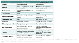
This needs to be distinguished from an omphalocoele, which is another type of abdominal wall defect.

An omphalocole is a midline herniation of the intestines (and, sometimes, other organs)  through the umbilical ring.

A critical point is that in an omphalocele, the contents are covered by the peritoneum and amniotic membrane.

These features and additional information are summarised in the following table

GI: Paedatric Pathology Case 2

A newborn presents with respiratory distress soon after birth.

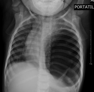
Chest X-ray reveals bowel loops in the left chest and mediastinal shift.

https://radiopaedia.org/articles/bochdalek-hernia


Congenital diaphragmatic hernia  (Bochdalek hernia – see below)

This is caused by the failure of the pleuroperitoneal membrane to fuse


Classification of Diaphragmatic Hernias

Category Type Subtype Key Features
Congenital  Bochdalek Hernia ~90% of Congenital Diaphragmatic Hernias ; postero-lateral defect; predominantly left-sided; impairs fetal lung development (pulmonary hypoplasia)
Congenital  Morgagni Hernia Anterior defect; rarely detected antenatally; less severe respiratory impact in infants
Congenital  Central Tendon Defect Very rare; defect in the central tendon of the diaphragm
Acquired Traumatic Blunt force (e.g. RTA) or penetrating injury (e.g. stab wound) causing diaphragmatic rupture
Acquired Hiatal Hernia Sliding (Type I) GOJ and stomach slide superiorly through the oesophageal hiatus; most common type
Acquired Hiatal Hernia Paraesophageal (Types II–IV) GOJ remains in situ; portion of stomach herniates alongside oesophagus; Types III/IV involve increasing gastric herniation
Acquired Iatrogenic Post-surgical diaphragmatic defect, typically following thoracic or upper abdominal procedures (e.g. oesophagectomy, fundoplication)


Robbin’s: Page 686


GI: Paedatric Pathology Case 1

A newborn presents with choking, coughing, and vomiting after taking the first feed. The nasogastric tube has passed but coils in the upper oesophagus.

The most likely diagnosis is oesophageal atresia, which is usually associated with a tracheoesophageal fistula.

Atresia, stenosis, fistulae, and duplications

These can occur anywhere in the gastrointestinal tract.

  1. Atresia is when there is a thin, non-canalised cord-like structure that replaces the normal structure.
  2. Stenosis is when the bowel is markedly narrowed. These are most clinically significant in the oesophagus and small intestine because of their relatively narrow lumen.
  3. A fistula is an abnormal communication between two epithelial-lined surfaces. The oesophagus and the trachea or bronchus in the examples below.
  4. Duplications are segments of the bowel that are duplicated!

Oesophageal atresia,

In the context of the oesophagus, atresia is very frequently associated with a tracheoesophageal fistula (and vice versa). There are a number of possibilities, with type C being the commonest.

en.wikipedia.org/…/Tracheoesophageal_fistula

Because there is a complete obstruction in the oesophagus immediately after birth, there are symptoms such as choking, coughing, and vomiting as a result of the obstruction, regurgitation and aspiration.

It should be noted that before birth, oesophageal atresia is associated with oligohydramnios

When an attempt is made to pass an oesophageal probe, it will not pass due to complete obstruction, which can be demonstrated on imaging.

Treatment is urgent surgery.

As usual, it is important to remember that once a congenital abnormality is present, there are likely to be others. Oesophageal atresia is, e.g., associated with an increased risk of imperforate anus.

In addition to atresia, congenital stenosis can occur. Other causes  (in adults)  of oesophageal stenosis include: gastroesophageal reflux, swallowing sodium hydroxide, radiation or scleroderma. Scleroderma and radiation also cause fibrosis in the lungs and heart (leading to cardiomyopathy).

Robbins:  page 685.

A plain-language guide to the molecular pathways in colorectal cancer

Colon Cancer is not one disease — it is several different diseases that all look similar under the microscope, but got there by very different routes. Understanding these routes helps explain why some bowel cancers behave differently, respond to different treatments, and arise in different patients.
There are three main roads to bowel cancer.

Road 1 — The Classical Route (the most common)
This is the familiar “polyp to cancer” story. A conventional polyp (adenoma) develops in the lining of the bowel and, over 10–15 years, accumulates a series of faults in key genes — first APC, then KRAS, then TP53. Each fault gives the cells a further growth advantage until cancer develops.
These cancers tend to be left-sided (descending colon and rectum) and are the ones most commonly detected by bowel cancer screening programmes.

Road 2 — The Serrated Route
This route starts from a different type of polyp — the sessile serrated lesion. These polyps are flat, pale, and easy to miss at colonoscopy, which is one reason this pathway is clinically important.
The first genetic fault here is in a gene called BRAF. What happens next is unusual — instead of accumulating more gene mutations, the tumour takes an epigenetic shortcut.
What does epigenetic mean?
Think of genes as light switches. A genetic mutation breaks the switch permanently. An epigenetic change is different — it puts a piece of “sticky tape” over the switch, keeping it off without actually breaking it. The gene is still physically intact but cannot be read. This chemical silencing process uses methyl groups and is called methylation.
In the serrated pathway, sticky tape is applied to the control switches of multiple genes simultaneously — a process called CIMP (CpG island methylator phenotype), or simply being “methylated”.
What happens next depends on which genes get silenced:
• If the sticky tape lands on MLH1 — the gene that proofreads and repairs DNA copying errors — the cell loses its ability to correct mistakes. Errors pile up rapidly in short repetitive DNA sequences called microsatellites. This is called microsatellite instability (MSI-H). These cancers have a very high number of mutations, attract a large immune response, tend to be right-sided, occur more often in older women, and respond very well to modern immunotherapy drugs.
• If the sticky tape does NOT silence MLH1, the cancer remains genomically stable (MSS). Paradoxically, this subgroup tends to behave more aggressively and does not respond to immunotherapy.
What about MSI-Low (MSI-L)?
You will sometimes see a third category — MSI-Low (MSI-L) — in reports and papers. This means there is a small degree of microsatellite instability, more than a fully stable tumour but nowhere near the level seen in MSI-H. In practice, MSI-L is a borderline or intermediate result. Most MSI-L tumours behave biologically more like MSS cancers than MSI-H cancers — they do not carry the same good prognosis, do not respond to immunotherapy, and are not generally associated with Lynch syndrome. MSI-L is thought to reflect minor, incidental errors in DNA copying rather than a true failure of the mismatch repair system. In most clinical algorithms, MSI-L is grouped with MSS for treatment decisions.

Road 3 — Lynch Syndrome (the inherited route)
Lynch syndrome also produces MSI-H cancers, but through a completely different mechanism. Here, a person is born with a faulty copy of one of the DNA repair genes. There is no sticky tape involved — the problem is hardwired into every cell of the body from conception.
How is it inherited?
Lynch syndrome follows an autosomal dominant pattern of inheritance. This means:
• Only one faulty copy of the gene is needed — inherited from one parent — to carry the syndrome. You do not need two faulty copies.
• Each child of an affected parent has a 50% chance of inheriting the faulty gene.
• It affects men and women equally.
• The faulty gene may have come from either the mother’s or father’s side of the family.
The genes involved are the mismatch repair genes: MLH1, MSH2, MSH6, and PMS2. Carrying a faulty copy does not guarantee cancer — it raises the lifetime risk substantially (up to 70–80% for bowel cancer with MLH1/MSH2 mutations) but is not inevitable. This is why Lynch families are offered regular surveillance colonoscopy.
Lynch syndrome also increases the risk of cancers in other organs — particularly the womb (endometrium), ovary, stomach, urinary tract, and small bowel — because the same repair machinery operates throughout the body.
The key distinction from the serrated pathway is that CIMP and BRAF mutation are absent in Lynch syndrome. Both arrive at MSI-H by different roads.

Detecting MMR Deficiency: IHC versus Molecular MSI Testing
In routine clinical practice, there are two completely different laboratory methods used to detect defective DNA repair in bowel cancers. They measure different things, and it is important not to confuse them.
Immunohistochemistry (IHC) — looking at the proteins
IHC is a staining technique performed by the pathologist on the tumour tissue section. Antibodies are applied that specifically bind to the four MMR proteins — MLH1, MSH2, MSH6, and PMS2 — and a colour reaction shows whether each protein is present or absent in the tumour cell nuclei.
In a normal tumour, all four proteins stain positively (present). If one or more proteins are lost, this indicates that the corresponding gene has been switched off or mutated — the cancer is said to show loss of MMR protein expression, or to be dMMR (deficient mismatch repair).
IHC has additional diagnostic value: the pattern of loss points towards the likely cause:
• Loss of MLH1 and PMS2 together → most likely sporadic (epigenetic silencing via CIMP, as in the serrated pathway). MLH1 methylation testing can confirm this.
• Loss of MSH2 and MSH6 together → strongly suggests Lynch syndrome (germline MSH2 mutation).
• Isolated loss of MSH6 or PMS2 → may indicate Lynch syndrome with a mutation in that specific gene.
• Loss of MLH1/PMS2 in a younger patient, or without BRAF mutation → raises suspicion for Lynch syndrome even if MLH1 is lost.
IHC is widely available, inexpensive, fast, and gives results the pathologist can interpret directly from the slide. However, it tests for protein — it tells you the protein is missing but does not directly measure what is happening to the DNA.
Molecular MSI Testing — looking at the DNA directly
Molecular MSI testing (also called PCR-based MSI testing or next-generation sequencing MSI analysis) works at the DNA level. It directly measures the lengths of specific short repetitive DNA sequences — microsatellites — in the tumour compared to normal tissue. If these sequences are abnormally variable in length, the tumour is called MSI-H. If they are stable, it is called MSS.
This test does not look at proteins at all. It confirms functionally that the DNA repair machinery has failed, regardless of which protein caused the problem or why.
So which test does what?
Think of it this way:
• IHC asks: which MMR protein is missing from the tumour? It identifies the defective component and points towards the mechanism (sporadic vs Lynch).
• Molecular MSI testing asks: has the loss of that protein actually caused DNA repair failure? It confirms the functional consequence.
• The two tests usually agree — a tumour that is dMMR by IHC is almost always MSI-H by molecular testing, and vice versa. But there are occasional discordant cases where one test is positive and the other negative, which is why both may be used in complex or equivocal situations.
• IHC is the standard first-line test in most pathology laboratories because it is practical and gives mechanistic clues. Molecular MSI testing is used to confirm, for clinical trials, or when IHC results are equivocal.
• Neither test on its own tells you whether the MMR deficiency is due to Lynch syndrome (germline, inherited) or sporadic methylation — that question requires germline genetic testing of the patient’s blood DNA, ideally guided by the IHC pattern and MLH1 methylation status.

The Bottom Line
• MSI (microsatellite instability) is the consequence — the end result of broken DNA repair, whatever the cause.
• MSI-H means the repair system has truly failed. MSI-L is a borderline finding that usually behaves like MSS and does not indicate Lynch syndrome.
• CIMP (methylation) is one mechanism that can cause MSI-H — by chemically silencing the MLH1 repair gene with sticky tape.
• The serrated pathway is the route — a distinct biological journey, starting from a different polyp, that frequently leads to CIMP and MSI-H.
• Lynch syndrome arrives at the same MSI-H destination by an entirely different, inherited road — a germline gene fault passed from parent to child with 50% probability.
• IHC and molecular MSI testing are complementary tools: IHC identifies which protein is lost and guides the search for the cause; molecular testing confirms that DNA repair has functionally failed.

Quick Reference Glossary
CIMP — CpG island methylator phenotype. The process of chemically silencing multiple genes simultaneously via methylation.
dMMR — Deficient mismatch repair. Detected by IHC as loss of one or more MMR proteins.
Epigenetic — Changes that affect gene activity without altering the DNA sequence itself.
IHC (Immunohistochemistry) — A staining technique that detects specific proteins in tissue sections using antibodies.
Methylation — Addition of methyl groups to gene promoters, acting like sticky tape over a switch to silence gene expression.
MMR — Mismatch repair. The cellular machinery that proofreads and corrects DNA copying errors.
MSI-H — Microsatellite instability-high. Confirms functional failure of DNA mismatch repair; high mutation burden; responds to immunotherapy.
MSI-L — Microsatellite instability-low. A borderline finding; behaves clinically like MSS; not associated with Lynch syndrome.
MSS — Microsatellite stable. Normal DNA repair function.
Autosomal dominant — Inheritance pattern where one faulty copy of a gene (from either parent) is sufficient to cause the condition; 50% transmission risk per child.
Lynch syndrome — An inherited condition caused by a germline mutation in an MMR gene (MLH1, MSH2, MSH6, or PMS2), predisposing to bowel and other cancers.
BRAF / KRAS / APC / TP53 — Genes that when mutated drive cancer development along different pathways.

Reference: Guinney et al., Nature Medicine 2015 (Consensus Molecular Subtypes) | WHO Classification of Digestive System Tumours

Duodenal biopsy site for diagnosing coeliac disease: D1, D2, or both?

Summary

Current best practice, in line with BSG, ESPGHAN and ACG guidance, is to take biopsies from both the duodenal bulb (D1) and the distal duodenum (D2 / D3), placed in separate pots. At least one (preferably two) bulb biopsies plus four or more distal duodenal biopsies is the standard recommendation. D2 remains the principal diagnostic site, but adding D1 increases diagnostic sensitivity, particularly for ultra-short coeliac disease and paediatric cases.

Historical position: D2 only

Traditional teaching was to biopsy the second part of the duodenum and to avoid the bulb. The rationale was that the bulb is architecturally complex — Brunner glands distort villous architecture, peptic injury and gastric metaplasia are common, and orientation in the histology laboratory is often poor. These features were felt to give unreliable assessment of villous atrophy and to risk false-positive interpretations of Marsh 1–3 change.

Why D1 was added: ultra-short coeliac disease

A series of studies in the 2000s and 2010s showed that a meaningful minority of coeliac patients have disease that is confined to, or most marked in, the bulb. Bonamico and colleagues demonstrated that taking bulb biopsies in addition to distal duodenum increased the diagnostic yield in children, and identified the 9 o’clock and 12 o’clock positions as the most informative bulb sites. Evans et al. and subsequent adult series have confirmed that around 5–10% of adult coeliac patients have lesions limited to or more severe in the bulb (ultra-short coeliac disease), and that omitting D1 would miss these cases.

Practical protocol

  • At least 1–2 biopsies from the duodenal bulb (ideally from the 9 o’clock and 12 o’clock positions, per Bonamico).
  • At least 4 biopsies from the distal duodenum (D2 / D3).
  • Bulb biopsies should be submitted in a separate, clearly labelled pot so the pathologist can apply appropriate caution when interpreting villous architecture around Brunner glands.
  • Single-biopsy-per-pass technique is preferred for orientation; multiple biopsies in one bite increase tangential sectioning and false-positive villous blunting.

Caveats with bulb biopsies

Bulb mucosa is intrinsically harder to assess. Brunner gland lobules push villi apart and shorten them, gastric metaplasia is common, and peptic duodenitis can produce IEL increases unrelated to gluten. Bulb-only abnormalities should therefore be interpreted in the context of serology, HLA status, and the distal duodenal appearances before a confident coeliac diagnosis is made.

Bottom line

Biopsy both. D2 remains the principal diagnostic site, but the addition of separately-potted bulb biopsies materially improves sensitivity, particularly for ultra-short coeliac disease and in children. This is the position of the BSG, ESPGHAN and ACG guidelines.

Key references

  • Ludvigsson JF, Bai JC, Biagi F, et al. Diagnosis and management of adult coeliac disease: guidelines from the British Society of Gastroenterology. Gut 2014;63:1210–1228.
  • Husby S, Koletzko S, Korponay-Szabó I, et al. European Society Paediatric Gastroenterology, Hepatology and Nutrition Guidelines for Diagnosing Coeliac Disease 2020. J Pediatr Gastroenterol Nutr 2020;70:141–156.
  • Rubio-Tapia A, Hill ID, Kelly CP, et al. ACG Clinical Guidelines: Diagnosis and Management of Celiac Disease. Am J Gastroenterol 2013;108:656–676 (and updated 2023 ACG guideline, Am J Gastroenterol 2023;118:59–76).
  • Bonamico M, Mariani P, Thanasi E, et al. Patchy villous atrophy of the duodenum in childhood celiac disease. J Pediatr Gastroenterol Nutr 2004;38:204–207.
  • Bonamico M, Thanasi E, Mariani P, et al. Duodenal bulb biopsies in celiac disease: a multicenter study. J Pediatr Gastroenterol Nutr 2008;47:618–622.
  • Evans KE, Aziz I, Cross SS, et al. A prospective study of duodenal bulb biopsy in newly diagnosed and established adult celiac disease. Am J Gastroenterol 2011;106:1837–1842.
  • Mooney PD, Kurien M, Evans KE, et al. Clinical and immunologic features of ultra-short celiac disease. Gastroenterology 2016;150:1125–1134.
  • Lebwohl B, Kapel RC, Neugut AI, et al. Adherence to biopsy guidelines increases celiac disease diagnosis. Gastrointest Endosc 2011;74:103–109.
  • Latorre M, Lagana SM, Freedberg DE, et al. Endoscopic biopsy technique in the diagnosis of celiac disease: one bite or two? Gastrointest Endosc 2015;81:1228–1233.

Cholangiocarcinoma

https://www.aasld.org/liver-fellow-network/core-series/why-series/why-do-we-transplant-some-types-cholangiocarcinoma-and

Cholangiocarcinoma (CCA) represents a heterogeneous group of malignancies arising from the biliary epithelium. It is divided into three subtypes depending on their anatomical site of origin:

intrahepatic (iCCA),

perihilar (pCCA) and

distal (dCCA).

iCCAs arise above the second-order bile ducts, while pCCA (also called a Klatskin tumor) arise above the cystic duct with dCCA coming from below the cystic duct.

pCCA is the single largest group, accounting for approximately 50–60% of all CCAs, followed by dCCA (20–30%) and iCCA (10–20%).

In this post, we will primarily discuss pCCA and iCCA, as dCCA is treated surgically with pancreaticoduodenectomy (Whipple procedure) rather than liver transplantation.

CCA accounts for approximately 3% of all gastrointestinal cancers representing the second most common primary hepatic malignancy after hepatocellular carcinoma (HCC).

Unlike HCC, the majority of CCA cases occur in the absence of an evident chronic liver disease or other risk factor making it more difficult to catch early.

Primary risk factors are Primary Sclerosing Cholangitis (PSC), with a lifetime risk of 10%, rising to 30% in those with Inflammatory Bowel Disease (which is why we annually screen these patients with an MRI), biliary cystic disease, and less commonly hepatitis B. Outside of the United States, southeast Asian liver flukes remain a risk factor.