Category: Hepato-biliary Pathology

BSEP (Bile Salt Export Pump) Immunohistochemistry

Bile Salt Export Pump (BSEP) is an ATP-binding cassette transporter protein (encoded by ABCB11) that functions as the major canalicular bile salt transporter in hepatocytes. In diagnostic pathology, BSEP IHC is used primarily as a canalicular marker for hepatocellular carcinoma and in the diagnosis of bile salt export pump deficiency in progressive familial intrahepatic cholestasis type 2 (PFIC2).

Hepatocellular carcinoma

BSEP shows a canalicular (“chicken-wire”) staining pattern in tumour cells, reflecting its normal physiological role as a bile salt transporter. This pattern confirms hepatocellular differentiation and helps distinguish HCC from metastatic adenocarcinoma or cholangiocarcinoma, neither of which forms canaliculi and therefore won’t show this staining pattern.

BSEP functions conceptually alongside other canalicular markers such as polyclonal CEA and CD10, and is generally considered more sensitive and specific than pCEA in some series.

Loss or reduction of BSEP staining has also been reported in poorly differentiated HCC and in some cholestatic hepatocellular lesions, so absent staining isn’t specific on its own — pattern interpretation in context matters.

Progressive Familial Intrahepatic Cholestasis Type 2

PFIC2 results from biallelic ABCB11 mutations. Liver biopsy IHC typically shows absent or markedly reduced canalicular BSEP staining, supporting the diagnosis alongside clinical/biochemical features (low-GGT cholestasis, elevated bile acids) and molecular confirmation.

A useful nuance: some ABCB11 missense mutations produce a mistrafficked but partially functional protein, which can show weak/patchy canalicular staining or an intracellular rather than purely canalicular pattern — so BSEP IHC is supportive but not strictly binary, and genetic testing remains the gold standard.

PFIC2 patients with BSEP deficiency also carry an increased risk of hepatocellular carcinoma and cholangiocarcinoma at a young age, which is relevant when assessing paediatric explant or resection specimens.
Cholangiocarcinoma

https://www.aasld.org/liver-fellow-network/core-series/why-series/why-do-we-transplant-some-types-cholangiocarcinoma-and

Cholangiocarcinoma (CCA) represents a heterogeneous group of malignancies arising from the biliary epithelium. It is divided into three subtypes depending on their anatomical site of origin:

intrahepatic (iCCA),

perihilar (pCCA) and

distal (dCCA).

iCCAs arise above the second-order bile ducts, while pCCA (also called a Klatskin tumor) arise above the cystic duct with dCCA coming from below the cystic duct.

pCCA is the single largest group, accounting for approximately 50–60% of all CCAs, followed by dCCA (20–30%) and iCCA (10–20%).

In this post, we will primarily discuss pCCA and iCCA, as dCCA is treated surgically with pancreaticoduodenectomy (Whipple procedure) rather than liver transplantation.

CCA accounts for approximately 3% of all gastrointestinal cancers representing the second most common primary hepatic malignancy after hepatocellular carcinoma (HCC).

Unlike HCC, the majority of CCA cases occur in the absence of an evident chronic liver disease or other risk factor making it more difficult to catch early.

Primary risk factors are Primary Sclerosing Cholangitis (PSC), with a lifetime risk of 10%, rising to 30% in those with Inflammatory Bowel Disease (which is why we annually screen these patients with an MRI), biliary cystic disease, and less commonly hepatitis B. Outside of the United States, southeast Asian liver flukes remain a risk factor.

Some Notes on Liver Disease

This was tricky for me to write as the subject is  the closest to my heart! I  really found it hard to leave so much out.


Acute hepatitis:

  1. Viruses
  2. Drugs


Chronic hepatitis (liver disease lasting more than 6 months)

  1. Viruses
  2. Autoimmune
  3. Drugs


Pathological features of chronic hepatitis

Grade = inflammation (can be in 3 places: portal tracts, interface and lobular)

Stage = fibrosis (portal tract expansion > bridging > cirrhosis)


The commonest causes of cirrhosis are:

  1. viral hepatitis
  2. alcoholic liver disease
  3. non-alcoholic fatty liver disease


Viral hepatitis

There are 5 hepatitis viruses (all RNA except for HBV): A-E

A and E: spread by faecal oral route, cause only an acute hepatitis

B, C and D (which can only infect people who have HBV, as well): spread by blood etc., cause the full range of liver disease:

  1. acute hepatitis
  2. chronic hepatitis – scarring begins
  3. cirrhosis – nodules of hepatocytes surrounded by scar tissue


Alcoholic liver disease and non-alcoholic fatty disease (risk factors: obesity, diabetes) produce the same pathological changes:

  1. fatty change
  2. fatty liver hepatitis (alcoholic hepatitis / non-alcoholic steatohepatitis (NASH) respectively): ballooning, neutrophils and scarring
  3. cirrhosis

NB These 3 stages often con-exist


There are (many) other liver diseases which may  cause of cirrhosis:

  1. Autoimmune hepatitis: anti smooth muscle actin autoantibodies, plasma cells, associated with other AI diseases, response to steroids
  2. Drug induced liver injury (DILI): “any kind of liver disease can be caused by a drug’
  3. Haemochromatosis = genetic (AR) increased iron absorption from the gut

deposited in the liver and many other organs (including the pancreas).

  1. Wilson’s disease = genetic (AR) decreased copper excretion (by hepatocytes into the bile duct).
  2. Primary sclerosing cholangitis (PSC): sclerosis (= fibrosis) of the bile ducts leading to their loss. Associated with Ulcerative Colitis
  3. Primary biliary cholangitis (PBC): inflammation (with granulomas) of the bile ducts leading to their loss


Complications of cirrhosis:

  1. Portal hypertension with varices
  2. Liver failure with hepatic encephalopathy,
  3. Liver cell cancer (the same as hepatocellular carcinoma)


Tumours of the liver:

The commonest are secondary tumours (many via the portal vein)


Primary tumours

  1. Benign
  2. Bile duct adenomas
  3. Hepatic adenomas (associated with the contraceptive pill)


  1. Malignant
  2. Liver cell carcinoma

Most commonly associated with cirrhosis.

Spread via the portal vein.

Carry a poor prognosis.


  1. Cholangiocarcinoma (an adenocarcinoma)

Divided into intrahepatic and extra hepatic (including gall bladder)

May be associated with ulcerative colitis and worm infections

Spread to lymph nodes

Carry a poor prognosis


Not “Everyone Must Get Stoned” *

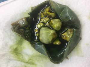
My first blog is going to be about gallstones. There is no special reason for this other than they are common (and, therefore, I hope this will be of interest to lots of people) and I happen to find gallbladder pathology interesting!

Below is a picture of a case I cut up yesterday:

As a medical student, I remember that the risk factors for gallstones were described as being “fat, fair, female and forty/ fertile ”  (depending on the specific version of the mnemonic).

How does this shape up now?  In this blog my core reference is Robin’s Pathology (in its range of formats) but will use a range of others. I will return to the topic of textbooks another time.

According to Robbins Basic Pathology (page 673):

Age: The prevalence of gallstones increases with age; over a 1/4 of people aged over 80 years have stones.

Sex: At every age, they are twice as common in women.

Ethnicity: They are very common in certain Native American groups.

Hereditary:  Family history and genetic disorders of bile salt metabolism. Although not mentioned in Robbins,  patients with haemolytic anaemias, including genetic ones such as Sickle Cell Anaemia, are at increased risk of pigment stones.

Environment:  Any factor that increases cholesterol excretion will increase the risk of stones. Oestrogens do this  (obviously contributing to the increased risk in women) as does obesity, rapid weight loss and drugs which increase cholesterol excretion, such as clofibrate.

Disorders affecting gallbladder motility: These includes pregnancy which contributes to the female and fertile risk.

So how does the mnemonic stand up? I came  across a paper (Postgrad Med J. 2013 Nov;89(1057):638-41. ) which directly addresses this question and concluded: “Our study found that the validated ‘students’ 5Fs’ mnemonic retains a role in clinical diagnosis of patients suspected of cholelithiasis but the factor ‘familial’ should be substituted for ‘forty’ in recognition of the role of inheritance and the changing demographics of gallstone incidence.”.

* Bob Dylan:  https://www.youtube.com/watch?v=ASQ-yHWKSQk